Parts of Your Chest Parts of Your Chest
Parts of Your Chest
The chest is one of the most critical areas of the human body, encompassing a wide range of anatomical structures that are vital for both movement and survival. When we talk about the "parts of your chest," we refer to everything from the external features like muscles and bones to the internal components such as organs and tissues. Understanding these parts can provide valuable insights into how our bodies function and why this region deserves special attention.
The chest region is not just a collection of random structures; it is an intricate system designed to protect and support some of the most essential functions of life. For instance, the ribcage shields delicate organs like the heart and lungs, while the muscles enable movement and strength. By exploring the various components of the chest, we gain a deeper appreciation for its complexity and importance in maintaining overall health.
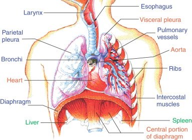
Let us delve further into the anatomy of the chest by examining its key structures. The chest is composed of bones, muscles, and internal organs, each playing a unique role. The sternum, or breastbone, serves as the central anchor point for the ribs, forming the front part of the ribcage. Meanwhile, the ribs and costal cartilages create a protective cage around vital organs. Beneath these protective layers lie the pectoralis major and minor muscles, which contribute significantly to upper body strength. Inside, the heart pumps blood through the circulatory system, and the lungs facilitate the exchange of oxygen and carbon dioxide during respiration. Together, these elements form a harmonious system that supports life itself.
The Chest Region
To better understand the chest, let's first focus on its broader context: the chest region. This area extends from the base of the neck down to the diaphragm, encompassing a large portion of the torso. It houses many important structures, making it one of the most dynamic and functional parts of the human body. The chest region is not only responsible for protecting vital organs but also plays a significant role in movement, posture, and even emotional expression.
The chest region is supported by a framework of bones, including the sternum, ribs, and spine. These bones work together to provide structural integrity and protection. Additionally, the chest contains several muscle groups, such as the pectoralis major and minor, which allow for a wide range of movements, from lifting objects to pushing against resistance. Even simple actions like breathing involve the coordinated effort of muscles in the chest region.
When discussing the chest region, it is important to recognize its dual roles: protective and functional. On one hand, the ribcage shields the heart and lungs from injury. On the other hand, the muscles enable movement and strength, contributing to daily activities like walking, running, and lifting. This balance between protection and functionality highlights the chest's versatility and importance in human anatomy.
Anatomical Structures
Within the chest region, numerous anatomical structures work together to ensure proper function. These structures can be broadly categorized into three groups: bones, muscles, and internal organs. Each group plays a specific role in maintaining the health and well-being of the chest.
Starting with the bones, the sternum serves as the central anchor point for the ribcage. It is a flat bone located at the front center of the chest, connecting the ribs via costal cartilages. The ribs themselves wrap around the torso, forming a protective cage for the internal organs. Together, these bones create a sturdy framework that safeguards the heart and lungs while allowing for flexibility during movement.
Moving on to the muscles, the chest contains two primary muscle groups: the pectoralis major and minor. The pectoralis major is a large, fan-shaped muscle that covers much of the chest region. It is responsible for powerful movements such as pushing and lifting. Beneath it lies the pectoralis minor, a smaller muscle that assists in shoulder movement and stabilization. Both muscles are crucial for upper body strength and contribute to the overall appearance of the chest.
Finally, the internal organs housed within the chest include the heart and lungs. The heart, a muscular organ, pumps blood throughout the body, delivering oxygen and nutrients to cells. The lungs, meanwhile, facilitate respiration by exchanging oxygen and carbon dioxide. These organs rely on the protective structure of the ribcage and the supportive function of the surrounding muscles to operate effectively.
The Sternum
At the core of the chest lies the sternum, often referred to as the breastbone. This flat, elongated bone runs vertically down the center of the chest and serves as the keystone of the ribcage. Its primary function is to connect the ribs via costal cartilages, forming a protective shield for the heart and lungs. Without the sternum, the ribcage would lack stability, leaving these vital organs vulnerable to injury.
The sternum consists of three main parts: the manubrium, body, and xiphoid process. The manubrium is the uppermost section, where the clavicles (collarbones) attach. Below it lies the body, the longest portion of the sternum, which connects to the ribs. Finally, the xiphoid process is a small, pointed structure at the bottom of the sternum. While the xiphoid process may seem insignificant, it plays a role in muscle attachment and serves as a landmark for medical procedures.
In addition to its protective role, the sternum also contributes to the overall structure of the chest. It helps maintain the shape of the ribcage, ensuring that there is enough space for the lungs to expand during inhalation. Furthermore, the sternum provides attachment points for several muscles, including the pectoralis major and rectus abdominis. These attachments allow for coordinated movement and strength in the upper body.
Ribs and Costal Cartilages
Surrounding the sternum are the ribs, which form the lateral walls of the ribcage. There are twelve pairs of ribs in total, each attached to the thoracic vertebrae in the back. The first seven pairs, known as true ribs, connect directly to the sternum via costal cartilages. The next three pairs, called false ribs, connect indirectly through the cartilage of the rib above them. Finally, the last two pairs are floating ribs, which do not attach to the sternum at all.
Costal cartilages play a crucial role in the flexibility of the ribcage. These bands of connective tissue link the ribs to the sternum, allowing for expansion and contraction during breathing. Unlike bones, cartilage is more flexible, enabling the ribcage to accommodate changes in volume as the lungs inflate and deflate. This elasticity is essential for efficient respiration and ensures that the chest can adapt to different levels of physical activity.
Together, the ribs and costal cartilages form a protective cage around the heart and lungs. This structure not only shields these vital organs from external forces but also provides a stable framework for muscle attachment. As a result, the ribcage supports both protection and movement, highlighting the intricate design of the chest region.
The Ribcage
The ribcage is a marvel of engineering, combining strength and flexibility to safeguard the internal organs of the chest. Composed of the sternum, ribs, and costal cartilages, the ribcage forms a bony enclosure that protects the heart and lungs while allowing for movement and expansion during respiration. Its design reflects the delicate balance between rigidity and adaptability required for optimal function.
One of the ribcage's most important roles is providing protection. The sturdy bones of the ribs and sternum create a barrier that shields the heart and lungs from impact and injury. This protective function is particularly crucial during physical activities or accidents, where the chest may be subjected to external forces. Without the ribcage, these vital organs would be far more susceptible to damage.
In addition to protection, the ribcage facilitates movement and flexibility. The costal cartilages connecting the ribs to the sternum allow for expansion and contraction during breathing. This elasticity enables the lungs to fill with air during inhalation and expel it during exhalation. Moreover, the ribcage supports the attachment of muscles involved in movement, such as the pectoralis major and serratus anterior. These muscles contribute to upper body strength and coordination.
Protecting Internal Organs
While the external anatomy of the chest focuses on bones and muscles, the internal anatomy emphasizes the vital organs it protects. Among these, the heart and lungs stand out as the most critical components. The heart, a muscular organ located slightly to the left of the sternum, pumps blood throughout the body, delivering oxygen and nutrients to cells. Its rhythmic contractions ensure that every part of the body receives the necessary resources to function properly.
Adjacent to the heart are the lungs, two spongy organs that facilitate the exchange of oxygen and carbon dioxide. During inhalation, air enters the lungs, where oxygen diffuses into the bloodstream. At the same time, carbon dioxide, a waste product of cellular metabolism, is removed from the blood and expelled during exhalation. This continuous cycle of gas exchange is essential for sustaining life.
The ribcage plays a pivotal role in protecting these internal organs. By forming a sturdy enclosure around the heart and lungs, it minimizes the risk of injury from external forces. Additionally, the flexibility provided by costal cartilages allows the ribcage to expand and contract during respiration, ensuring that the lungs have sufficient space to function effectively. This combination of protection and adaptability underscores the importance of the ribcage in maintaining the health of the chest region.
Pectoralis Major Muscle
Beneath the skin and ribs lies the pectoralis major muscle, one of the largest and most prominent muscles in the chest region. This fan-shaped muscle extends from the sternum and clavicle to the humerus, the long bone of the upper arm. Its primary function is to facilitate powerful movements such as pushing and lifting. Whether you're performing a bench press at the gym or simply pushing a door open, the pectoralis major is actively engaged.
The pectoralis major is divided into two heads: the clavicular head and the sternal head. The clavicular head originates from the medial half of the clavicle, while the sternal head arises from the sternum and the upper six costal cartilages. These distinct origins allow the muscle to generate force in multiple directions, enhancing its versatility. Together, the two heads converge onto the humerus, creating a strong connection that enables effective movement.
In addition to its functional role, the pectoralis major contributes to the aesthetic appearance of the chest. Well-developed pectoral muscles are often associated with strength and fitness, making them a popular target for exercise enthusiasts. Exercises like push-ups, bench presses, and flyes are specifically designed to strengthen and tone this muscle group, improving both physical performance and visual appeal.
Pectoralis Minor Muscle
Below the pectoralis major lies the pectoralis minor, a smaller yet equally important muscle in the chest region. This triangular muscle originates from the third, fourth, and fifth ribs and inserts into the coracoid process of the scapula, a bony projection near the shoulder blade. Its primary function is to assist in shoulder movement and stabilization.
The pectoralis minor plays a key role in protraction and depression of the scapula, which are essential for maintaining proper shoulder alignment. When activated, it pulls the scapula forward and downward, helping to position the shoulder joint correctly. This action is particularly important during activities that require overhead movement, such as throwing or swimming.
Although less visible than the pectoralis major, the pectoralis minor is vital for overall shoulder health. Imbalances or tightness in this muscle can lead to postural issues, such as rounded shoulders or forward head posture. Therefore, incorporating exercises that target the pectoralis minor, such as wall slides or scapular retractions, can help improve posture and prevent discomfort.
Movement and Strength
The movement and strength of the chest region are largely dependent on the muscles found there. The pectoralis major and minor, along with other supporting muscles, work together to enable a wide range of motions. These muscles not only contribute to physical performance but also play a role in maintaining good posture and preventing injury.
Strength training exercises are an effective way to enhance the function of chest muscles. Activities like bench presses, push-ups, and dumbbell flyes target the pectoralis major, increasing its size and power. Similarly, exercises that focus on scapular movement, such as rows and pull-downs, engage the pectoralis minor, promoting better shoulder alignment and stability. By incorporating a variety of exercises into your routine, you can ensure balanced development of the chest muscles.
Proper technique is crucial when performing chest exercises. Using incorrect form can lead to strain or injury, undermining the benefits of strength training. To maximize results while minimizing risks, it is important to start with lighter weights and gradually increase intensity as your muscles become stronger. Additionally, incorporating rest days into your routine allows for recovery and growth, ensuring long-term progress.
The Heart
At the center of the chest lies the heart, a remarkable organ responsible for pumping blood throughout the body. Shaped like a cone and roughly the size of a fist, the heart is composed of specialized muscle tissue called cardiac muscle. This muscle contracts rhythmically, propelling blood through a network of arteries, veins, and capillaries. Every day, the heart beats approximately 100,000 times, circulating about 7,000 liters of blood.
The heart is divided into four chambers: the right and left atria, and the right and left ventricles. Blood enters the heart through the atria, which serve as receiving chambers. From there, it flows into the ventricles, which pump blood out to the rest of the body. The right side of the heart handles deoxygenated blood, sending it to the lungs for oxygenation, while the left side manages oxygen-rich blood, distributing it to tissues and organs.
Maintaining heart health is essential for overall well-being. Regular exercise, a balanced diet, and adequate rest are key factors in keeping the heart functioning optimally. Cardiovascular exercises, such as running, cycling, or swimming, strengthen the heart muscle and improve circulation. Avoiding harmful habits like smoking and excessive alcohol consumption also contributes to heart health, reducing the risk of cardiovascular disease.
Blood Circulation
The heart's primary function is to facilitate blood circulation, ensuring that every cell in the body receives the oxygen and nutrients it needs to survive. This process begins in the right atrium, where deoxygenated blood from the body enters the heart. It then flows into the right ventricle, which pumps the blood to the lungs for oxygenation. In the lungs, carbon dioxide is removed, and fresh oxygen is absorbed into the bloodstream.
Once oxygenated, the blood returns to the heart via the pulmonary veins, entering the left atrium. From there, it moves into the left ventricle, the most powerful chamber of the heart. The left ventricle contracts forcefully, sending oxygen-rich blood through the aorta and into the systemic circulation. This network of arteries, veins, and capillaries delivers blood to every part of the body, supporting cellular function and maintaining homeostasis.
Proper blood circulation is vital for maintaining health. Conditions that impede circulation, such as atherosclerosis or hypertension, can lead to serious complications, including heart attacks and strokes. To promote healthy circulation, it is important to adopt lifestyle habits that support cardiovascular health. Regular physical activity, a nutritious diet, and stress management techniques can all contribute to improved circulation and reduced risk of cardiovascular disease.
The Lungs
Adjacent to the heart are the lungs, two sponge-like organs that play a crucial role in respiration. Located on either side of the chest cavity, the lungs expand and contract with each breath, facilitating the exchange of oxygen and carbon dioxide. Their structure and function are intricately linked, ensuring efficient gas exchange and supporting overall health.
Each lung is divided into lobes: the right lung has three lobes, while the left lung has two, accommodating the presence of the heart. Within the lungs, a complex network of airways leads to tiny sacs called alveoli, where gas exchange occurs. Oxygen from inhaled air diffuses into the bloodstream through the thin walls of the alveoli, while carbon dioxide, a waste product of cellular metabolism, is expelled during exhalation.
Respiratory health depends on maintaining clear airways and functioning lungs. Practices such as avoiding smoking, minimizing exposure to pollutants, and practicing deep breathing exercises can help preserve lung function. Additionally, regular aerobic exercise strengthens the respiratory muscles, improving lung capacity and efficiency. By prioritizing lung health, individuals can enhance their overall well-being and reduce the risk of respiratory diseases.
Respiration Process
The respiration process involves a series of steps that ensure the continuous exchange of gases between the body and the environment. It begins with inhalation, during which air enters the nose or mouth and travels down the trachea into the bronchi and bronchioles of the lungs. These airways branch repeatedly, eventually leading to the alveoli, where gas exchange occurs.
Inside the alveoli, oxygen diffuses into the bloodstream through capillaries, binding to hemoglobin in red blood cells. Simultaneously, carbon dioxide, a waste product of cellular metabolism, moves from the blood into the alveoli to be expelled during exhalation. This exchange of gases is facilitated by the thin walls of the alveoli and the rich supply of capillaries surrounding them.
Efficient respiration requires healthy lungs and clear airways. Conditions such as asthma, chronic obstructive pulmonary disease (COPD), or pneumonia can impair respiratory function, making it difficult to breathe. To maintain optimal respiratory health, it is important to avoid harmful behaviors and environments, such as smoking or exposure to pollutants. Engaging in regular exercise and practicing breathing techniques can also enhance lung function and promote overall well-being.
External Anatomy
When examining the chest, it is helpful to distinguish between its external anatomy and internal anatomy. The external anatomy includes the visible features of the chest, such as the skin, muscles, and bones. These structures form the outer layer of the chest region, providing both protection and aesthetic appeal.
The skin covering the chest is relatively thick and elastic, allowing for movement and flexibility. Beneath the skin lie the pectoralis major and minor muscles, which contribute to the contour and shape of the chest. The sternum and ribs form the bony framework of the chest, creating a protective barrier for the internal organs. Together, these external structures define the appearance and function of the chest region.
Understanding the external anatomy of the chest is important for recognizing potential issues. Changes in skin texture, muscle tone, or bone structure may indicate underlying health concerns. For example, dimpling or retraction of the skin could signal breast cancer, while asymmetry in the chest muscles might suggest muscular imbalances. By paying attention to these external signs, individuals can identify problems early and seek appropriate medical care.
Internal Anatomy
While the external anatomy focuses on visible features, the internal anatomy delves deeper into the structures hidden beneath the surface. These include the heart, lungs, and other vital organs housed within the chest cavity. The internal anatomy is responsible for sustaining life by regulating processes such as blood circulation and respiration.
The heart, located slightly to the left of the sternum, serves as the engine driving the circulatory system. Its rhythmic contractions propel blood through a network of vessels, delivering oxygen and nutrients to cells. Adjacent to the heart are the lungs, which facilitate the exchange of gases during respiration. Together, these organs ensure that the body receives the resources it needs to function effectively.
Protecting these internal structures is the ribcage, a bony enclosure formed by the sternum, ribs, and costal cartilages. This framework shields the heart and lungs from injury while allowing for movement and flexibility. The internal anatomy of the chest is a testament to the body's intricate design, balancing protection with functionality to support life.
Bones and Muscles
The bones and muscles of the chest work together to create a strong, flexible framework that supports movement and protects internal organs. The sternum, ribs, and costal cartilages form the bony structure of the chest, providing a stable foundation for muscle attachment. Meanwhile, the pectoralis major and minor muscles enable powerful movements, contributing to upper body strength and coordination.
The interaction between bones and muscles is essential for maintaining proper function. For example, the pectoralis major attaches to the sternum and ribs, allowing it to generate force during pushing and lifting movements. Similarly, the pectoralis minor connects to the ribs and scapula, assisting in shoulder stabilization and movement. This synergy between bones and muscles ensures that the chest region can perform a wide range of activities with precision and efficiency.
To optimize the function of bones and muscles, it is important to engage in regular exercise and maintain a balanced diet. Strength training exercises targeting the chest muscles can improve their size and power, while flexibility exercises like stretching can enhance range of motion. Adequate intake of calcium and vitamin D supports bone health, reducing the risk of fractures and osteoporosis. By prioritizing the health of bones and muscles, individuals can enhance their overall physical capabilities.
Organs and Tissues
Within the chest cavity lie several vital organs and tissues that sustain life. Foremost among these are the heart and lungs, which work in tandem to ensure proper circulation and respiration. Surrounding these organs are layers of tissue, including fat, connective tissue, and nerves, which provide support and insulation.
The heart, a muscular organ, pumps blood throughout the body, delivering oxygen and nutrients to cells. Its rhythmic contractions maintain a steady flow of blood, ensuring that every part of the body receives the resources it needs. Adjacent to the heart are the lungs, which facilitate the exchange of gases during respiration. Oxygen enters the bloodstream through the alveoli, while carbon dioxide is expelled during exhalation.
Supporting these organs are various tissues that contribute to their function and protection. Fat tissue cushions the heart and lungs, reducing friction and shock during movement. Connective tissue binds structures together, maintaining the integrity of the chest cavity. Nerves transmit signals between the brain and chest organs, regulating processes such as heart rate and breathing. Together, these tissues ensure the smooth operation of the chest's internal systems.
Complexity of the Chest
The complexity of the chest is evident in its diverse array of structures and functions. From the protective framework of the ribcage to the powerful muscles of the chest, every component plays a unique role in maintaining health and supporting life. This complexity is further highlighted by the intricate interactions between bones, muscles, and internal organs, all working together to ensure optimal performance.
To fully appreciate the complexity of the chest, it is important to consider its dual roles: protective and functional. The ribcage shields the heart and lungs from injury, while the muscles enable movement and strength. The heart and lungs, in turn, regulate critical processes such as blood circulation and respiration. This interdependence underscores the importance of maintaining the health of all chest structures.
A checklist for maintaining chest health might include the following steps:
- Engage in regular exercise: Incorporate strength training and cardiovascular exercises to strengthen chest muscles and improve circulation.
- Adopt a balanced diet: Ensure adequate intake of nutrients like calcium and vitamin D to support bone health.
- Practice deep breathing exercises: Enhance lung function and reduce stress through controlled breathing techniques.
- Avoid harmful habits: Minimize exposure to pollutants and refrain from smoking to protect respiratory health.
- Monitor for changes: Pay attention to any alterations in chest appearance or function, seeking medical advice if necessary.
By following these steps, individuals can preserve the health and functionality of their chest region, enjoying a higher quality of life.
Deja una respuesta