Parts of a Dog's Eye
Parts of a Dog's Eye
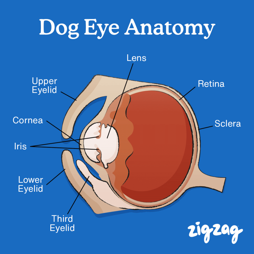
The dog's eye is an intricate and fascinating organ that plays a crucial role in its daily life. Like humans, dogs rely heavily on their vision to navigate the world around them. However, there are distinct differences in how their eyes function compared to ours. Understanding the anatomy of a dog's eye not only helps us appreciate this remarkable organ but also aids in recognizing potential health issues that may arise. In this section, we will delve into the various components of a dog's eye, starting with the cornea, which serves as the transparent outer layer.
Cornea
The cornea is one of the most vital parts of a dog's eye. It acts as the protective outermost layer while simultaneously allowing light to enter the eye. This clear, dome-shaped structure is responsible for focusing light onto the lens, enabling the dog to perceive objects clearly. The cornea is composed of several layers, including the epithelium, Bowman's layer, stroma, Descemet's membrane, and the endothelium. Each layer has a specific function, ensuring the cornea remains transparent and functional.
In addition to focusing light, the cornea protects the inner structures of the eye from external damage. Its smooth surface ensures that light enters the eye evenly, minimizing distortion. However, the cornea is also susceptible to injuries such as scratches or ulcers, which can impair vision if left untreated. Regular veterinary check-ups are essential to monitor the health of the cornea and detect any abnormalities early.
Interestingly, the cornea does not contain blood vessels, as these would interfere with its transparency. Instead, it relies on the tear film and aqueous humor for nourishment. Maintaining a healthy tear production system is crucial for keeping the cornea hydrated and functioning properly. Owners should pay close attention to any signs of redness, swelling, or discharge, as these could indicate corneal issues that require immediate attention.
Iris
Behind the cornea lies the iris, the colored part of the eye that gives it its distinctive appearance. The iris is responsible for controlling the size of the pupil, thereby regulating the amount of light that enters the eye. In bright conditions, the iris contracts the pupil to reduce light intake, while in dim environments, it expands the pupil to allow more light to pass through. This mechanism ensures optimal vision under varying lighting conditions.
The color of a dog's iris can vary widely depending on the breed and genetic makeup. Common colors include brown, blue, and amber, although some dogs may have heterochromia, where each eye has a different color. Despite these variations, the function of the iris remains consistent across all breeds. By adjusting the pupil size, the iris helps protect the sensitive retina from excessive light exposure, preventing potential damage.
It is important to note that changes in the appearance of the iris, such as cloudiness or discoloration, can be indicative of underlying health issues. Conditions like uveitis (inflammation of the iris) or cataracts can affect the clarity and function of the iris. Regular monitoring of the eyes, along with routine veterinary care, can help identify and address these problems promptly.
Pupil
The pupil is the opening at the center of the iris that allows light to enter the eye. Its size fluctuates based on the amount of light present in the environment, thanks to the muscles within the iris. When exposed to bright light, the pupil constricts to minimize glare and protect the delicate structures behind it. Conversely, in low-light situations, the pupil dilates to capture as much light as possible, enhancing the dog's ability to see in the dark.
This adaptive mechanism is crucial for a dog's survival, especially in the wild, where they often hunt during dawn and dusk when lighting conditions are less favorable. The rapid adjustment of the pupil ensures that the dog can quickly adapt to changing environments, maintaining clear vision regardless of the light levels.
However, abnormal pupil behavior, such as unequal sizes between the two eyes or a lack of response to light, can signal serious health concerns. These issues may stem from neurological disorders, trauma, or infections. If owners notice any irregularities in their dog's pupils, it is imperative to seek veterinary assistance immediately to prevent further complications.
Detailed Checklist for Monitoring the Cornea, Iris, and Pupil
To ensure your dog's eyes remain healthy, follow this detailed checklist:
Regular Eye Examinations: Conduct weekly inspections of your dog's eyes to check for any signs of redness, swelling, or discharge. Use a well-lit area to get a clear view of the cornea, iris, and pupil.
Observe Pupil Response: Shine a flashlight briefly into your dog's eyes to observe how the pupils react. They should constrict rapidly in response to light. If you notice sluggish or absent responses, consult a veterinarian.
Check for Corneal Issues: Look for cloudiness, scratches, or ulcers on the cornea. These could indicate injury or infection. Keep your dog's eyes clean and free of debris to prevent irritation.
Monitor Iris Color: Pay attention to any changes in the color or texture of the iris. Discoloration or cloudiness may point to underlying conditions like uveitis or cataracts.
Maintain Tear Production: Ensure your dog produces sufficient tears to keep the cornea hydrated. Dry eyes can lead to discomfort and potential damage. Consult a vet if you suspect tear production issues.
Lens
Positioned behind the iris, the lens is another critical component of the dog's eye. Its primary function is to focus light onto the retina, refining the image formed by the cornea. The lens achieves this through a process called accommodation, where it changes shape to adjust the focal length depending on the distance of the object being viewed. This flexibility allows dogs to see both near and far objects clearly.
As dogs age, the lens can lose some of its elasticity, leading to decreased flexibility and difficulty focusing on close objects. This condition, known as presbyopia, is similar to what humans experience as they grow older. Additionally, the lens can develop cloudiness due to cataracts, which significantly impair vision if left untreated. Early detection and management of cataracts are crucial to preserving the dog's sight.
Despite these potential challenges, the lens remains a vital part of the visual system, working in tandem with other ocular structures to provide clear and accurate vision. Owners should be vigilant about monitoring their dog's eyes for any signs of lens-related issues, such as cloudiness or difficulty navigating familiar environments.
Retina
At the back of the eye lies the retina, a thin layer of tissue that contains specialized cells called photoreceptors. These cells convert light into electrical signals, which are then transmitted to the brain via the optic nerve. The retina is essentially the "camera" of the eye, capturing images and sending them to the brain for interpretation. Without a healthy retina, vision would not be possible.
The retina is divided into two main regions: the central retina, which provides sharp, detailed vision, and the peripheral retina, responsible for detecting movement and providing a wider field of view. Dogs rely heavily on their peripheral vision, as it allows them to detect potential threats or prey from a distance. This evolutionary adaptation enhances their survival skills in the wild.
Certain conditions, such as retinal detachment or degeneration, can severely impact the retina's ability to function properly. Symptoms may include blindness, difficulty seeing in low light, or a reluctance to move in unfamiliar environments. Prompt diagnosis and treatment are essential to prevent permanent vision loss.
Rods and Cones
Within the retina are two types of photoreceptor cells: rods and cones. These cells play distinct roles in vision, with rods being responsible for detecting light and cones for perceiving color. Dogs have a higher concentration of rods than cones, which explains why they excel at seeing in low-light conditions but have limited color perception compared to humans.
Rods are highly sensitive to light, making them ideal for night vision. They enable dogs to detect even the faintest movements in dim environments, a trait that is particularly useful for nocturnal hunters. On the other hand, cones are responsible for color vision, although dogs possess fewer cone types than humans. As a result, their color perception is limited to shades of blue and yellow, meaning they cannot distinguish red or green hues.
Understanding the balance between rods and cones in a dog's retina highlights the unique way they perceive the world. While their color vision may not be as vibrant as ours, their enhanced sensitivity to light and motion compensates for this limitation, providing them with a distinct advantage in certain scenarios.
Tapetum Lucidum
One of the most fascinating features of a dog's eye is the tapetum lucidum, a reflective layer located behind the retina. This structure enhances night vision by reflecting light back through the retina, allowing photoreceptor cells a second chance to absorb it. This adaptation increases the efficiency of light detection, enabling dogs to see remarkably well in low-light conditions.
The tapetum lucidum is also responsible for the characteristic "eye shine" observed when light hits a dog's eyes at night. This phenomenon occurs because the reflective layer bounces light back toward the source, creating a glowing effect. The color of the shine can vary depending on the breed and individual dog, ranging from green to blue or yellow.
While the tapetum lucidum provides significant advantages in low-light environments, it can sometimes interfere with vision during the day. The reflection of light can cause glare, making it harder for dogs to see clearly in bright conditions. However, this drawback is outweighed by the benefits it offers in dimly lit situations, where vision is crucial for survival.
Sclera
Surrounding the cornea and other internal structures is the sclera, the tough, white outer layer of the eye. Its primary function is to provide structural support and protection to the delicate tissues within. The sclera is composed of dense connective tissue, making it strong and resilient against physical damage.
Although the sclera is not directly involved in the process of vision, it plays an essential role in maintaining the overall health and integrity of the eye. Any injury or inflammation affecting the sclera can lead to serious complications, including pain, redness, and impaired vision. Conditions such as scleritis, characterized by inflammation of the sclera, require prompt medical attention to prevent long-term damage.
Owners should regularly inspect their dog's eyes for signs of scleral issues, such as persistent redness or swelling. Keeping the eyes clean and free of irritants can help reduce the risk of infections or injuries that may affect the sclera.
Aqueous Humor and Vitreous Body
Two additional components that contribute to the health and function of a dog's eye are the aqueous humor and vitreous body. The aqueous humor is a clear fluid located in the anterior chamber of the eye, between the cornea and the lens. It plays a vital role in maintaining intraocular pressure and nourishing the cornea and lens by delivering essential nutrients and removing waste products.
On the other hand, the vitreous body is a gel-like substance that fills the posterior chamber of the eye, providing structural support and helping maintain its shape. It also serves as a medium for light to pass through, ensuring that images are focused accurately onto the retina. Over time, the vitreous body can deteriorate, leading to conditions such as floaters or retinal detachment.
Maintaining the health of both the aqueous humor and vitreous body is crucial for preserving vision. Regular veterinary check-ups can help detect any abnormalities early, allowing for timely intervention and treatment. Owners should be aware of symptoms such as cloudy eyes, squinting, or excessive tearing, which may indicate issues with these vital structures.
By understanding the intricate workings of a dog's eye, we gain a deeper appreciation for this remarkable organ and the importance of proper care and maintenance. Whether it's monitoring the cornea, iris, or any of the other components discussed, vigilance and proactive measures are key to ensuring lifelong ocular health for our canine companions.
Deja una respuesta