Parts of the Foot
Parts of the Foot
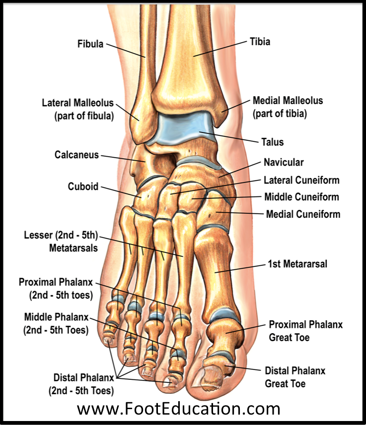
The human foot is a marvel of engineering, combining strength and flexibility to allow us to walk, run, jump, and maintain balance. Composed of 26 bones, numerous joints, muscles, tendons, and ligaments, the foot plays an essential role in our daily lives. Understanding its anatomy is not only fascinating but also crucial for diagnosing and treating various foot-related conditions. This section will delve into the intricate details of all parts of the foot, providing a comprehensive overview.
The foot can be divided into three main sections: the hindfoot, which includes the heel and ankle; the midfoot, characterized by the arches formed by specific bones; and the forefoot, consisting of the metatarsals and toes (phalanges). Each part has unique functions and structures that work together seamlessly to ensure optimal performance. Let’s explore these components in greater detail.
Hindfoot
The hindfoot is the posterior portion of the foot, encompassing the heel and ankle. It serves as the foundation for weight-bearing activities and provides stability during movement. The hindfoot is primarily responsible for shock absorption and transferring forces from the ground up through the leg.
Heel
The heel is the largest bone in the foot, known anatomically as the calcaneus. Its primary function is to absorb impact when the foot strikes the ground. The calcaneus is shaped like a wedge, allowing it to distribute weight evenly across the foot. Surrounding soft tissues, such as fat pads, cushion the heel and protect it from excessive pressure. Common conditions affecting the heel include plantar fasciitis and heel spurs, both of which can cause significant discomfort.
When walking or running, the heel acts as the initial point of contact with the ground. This makes it vulnerable to injuries, especially if proper footwear is not worn. To prevent heel pain, it is important to choose shoes with adequate cushioning and support. Additionally, stretching exercises targeting the Achilles tendon and calf muscles can help maintain flexibility and reduce strain on the heel.
Ankle
The ankle is a complex joint that connects the foot to the lower leg. It consists of three bones: the tibia (shinbone), fibula (calf bone), and talus (ankle bone). The ankle joint allows for dorsiflexion (lifting the foot upward) and plantarflexion (pointing the foot downward). These movements are critical for walking, running, and jumping.
Ligaments surrounding the ankle provide stability and prevent excessive motion that could lead to injury. The most commonly injured ligament in the ankle is the anterior talofibular ligament, often damaged during sprains. Strengthening the muscles around the ankle, particularly the peroneals and tibialis anterior, can enhance stability and reduce the risk of injury.
Proper alignment of the ankle joint is essential for maintaining balance and preventing long-term issues such as arthritis. Exercises that focus on proprioception, or the body's ability to sense its position in space, can improve ankle function and reduce the likelihood of falls.
Midfoot
The midfoot lies between the hindfoot and forefoot, forming the arches of the foot. These arches play a vital role in distributing weight and absorbing shock. The midfoot contains several small bones, including the navicular, cuboid, and cuneiform bones, which work together to create a stable yet flexible structure.
Arches
The foot has three main arches: the medial longitudinal arch, lateral longitudinal arch, and transverse arch. The medial longitudinal arch is the highest and most prominent, extending from the heel to the ball of the foot. It is supported by ligaments and tendons, including the plantar fascia, and helps elevate the foot during movement.
The lateral longitudinal arch is shorter and less pronounced than the medial arch. It runs along the outer edge of the foot and contributes to stability during weight-bearing activities. The transverse arch spans across the width of the foot, helping to maintain balance and distribute pressure evenly.
Flat feet, or fallen arches, occur when the arches collapse due to weakened ligaments or muscle imbalances. This condition can lead to pain and discomfort, particularly in the heels and ankles. Custom orthotics or supportive footwear may be recommended to alleviate symptoms and restore proper alignment.
Navicular Bone
The navicular bone is located on the inner side of the foot, near the ankle. It connects the talus (ankle bone) to the cuneiform bones and forms part of the medial longitudinal arch. The navicular bone plays a key role in stabilizing the foot and facilitating smooth movement.
Injuries to the navicular bone, such as fractures or stress fractures, are relatively common among athletes who engage in high-impact activities. Symptoms may include swelling, pain, and difficulty walking. Rest, immobilization, and physical therapy are often prescribed to promote healing and restore function.
Cuboid Bone
The cuboid bone is situated on the outer side of the foot, near the base of the fifth metatarsal. It forms part of the lateral longitudinal arch and works in conjunction with the calcaneus and cuneiform bones to provide stability. The cuboid bone also serves as an attachment point for several ligaments and tendons.
Cuboid syndrome, a condition characterized by pain and tenderness in the area, can result from overuse or improper foot mechanics. Treatment typically involves rest, ice, and manual manipulation to realign the bone. Strengthening exercises targeting the muscles of the foot and lower leg can help prevent recurrence.
Cuneiform Bones
The cuneiform bones are three small bones located in the midfoot, adjacent to the navicular bone. They connect the metatarsals to the navicular and cuboid bones, forming the medial and lateral longitudinal arches. Together, the cuneiform bones contribute to the overall stability and flexibility of the foot.
Disorders affecting the cuneiform bones, such as arthritis or fractures, can impair foot function and cause significant discomfort. Early diagnosis and treatment are essential for preserving mobility and preventing long-term complications. Non-surgical interventions, such as anti-inflammatory medications and custom orthotics, are often effective in managing symptoms.
Forefoot
The forefoot includes the metatarsals and toes (phalanges), which are responsible for propulsion during walking and running. This region of the foot is highly mobile, allowing for fine-tuned adjustments to terrain and surface variations.
Metatarsals
The metatarsals are five long bones that extend from the midfoot to the toes. They form the ball of the foot and play a crucial role in weight distribution and push-off during gait. Each metatarsal is numbered one through five, starting with the big toe.
Metatarsalgia, a condition characterized by pain and inflammation in the metatarsal region, is a common complaint among runners and dancers. Causes may include overuse, poor footwear, or structural abnormalities such as high arches or flat feet. Padding, arch supports, and strengthening exercises can help alleviate symptoms and improve foot health.
Toes
The toes are the terminal digits of the foot, consisting of three phalanges each (proximal, middle, and distal) except for the big toe, which has only two. They assist with balance, grip, and propulsion during movement. The toes are connected to the metatarsals by joints that allow for flexion and extension.
Conditions affecting the toes, such as bunions, hammertoes, and ingrown toenails, can significantly impact foot function and comfort. Proper nail care, wearing appropriately fitting shoes, and addressing underlying biomechanical issues can help prevent these problems. In severe cases, surgical intervention may be necessary to restore normal alignment and function.
Phalanges
The phalanges are the small bones that make up the toes. They are classified as short bones and are arranged in three segments: proximal, middle, and distal. The phalanges work in concert with the metatarsals and other structures to facilitate movement and provide stability.
Injuries to the phalanges, such as fractures or dislocations, are relatively common and can result from trauma or repetitive stress. Treatment depends on the severity of the injury and may involve splinting, casting, or surgical repair. Rehabilitation exercises focusing on range of motion and strength can aid in recovery and restore functionality.
Soft Tissues
In addition to bones, the foot contains various soft tissues that contribute to its overall function and integrity. These include the plantar fascia, Achilles tendon, flexor tendons, and extensor tendons. Each plays a distinct role in supporting the foot and enabling movement.
Plantar Fascia
The plantar fascia is a thick band of connective tissue that runs along the bottom of the foot, connecting the heel to the toes. It helps form the arches of the foot and provides shock absorption during weight-bearing activities. Stretching and strengthening exercises targeting the plantar fascia can help prevent conditions such as plantar fasciitis, which causes heel pain and stiffness.
Achilles Tendon
The Achilles tendon is the largest and strongest tendon in the body, connecting the calf muscles to the heel bone. It enables powerful movements such as pushing off during walking and running. Overuse or sudden increases in activity can lead to Achilles tendinitis, characterized by pain and swelling. Gradual progression of exercise intensity and incorporating eccentric strengthening exercises can reduce the risk of injury.
Flexor Tendons
The flexor tendons run along the underside of the foot and are responsible for bending the toes. They originate from muscles in the lower leg and insert into the phalanges. Damage to the flexor tendons can result in decreased toe flexion and impaired gait. Surgical repair may be required in cases of severe injury or rupture.
Extensor Tendons
The extensor tendons are located on the top of the foot and control toe extension. They arise from muscles in the front of the leg and attach to the phalanges. Conditions affecting the extensor tendons, such as tendonitis or tenosynovitis, can cause pain and swelling. Conservative treatments, including rest, ice, and anti-inflammatory medications, are usually sufficient for resolving symptoms.
Detailed Checklist for Maintaining Foot Health
To ensure optimal foot health and prevent common conditions, follow this detailed checklist:
Wear Proper Footwear: Choose shoes that fit well and provide adequate support. Look for features such as cushioned soles, arch support, and breathable materials. Replace worn-out shoes promptly to avoid unnecessary strain on the feet.
Stretch Regularly: Incorporate stretching exercises into your daily routine to maintain flexibility and reduce tension in the muscles and tendons of the foot. Focus on the Achilles tendon, calf muscles, and plantar fascia for best results.
Strengthen Foot Muscles: Perform exercises designed to strengthen the intrinsic muscles of the foot. Examples include toe curls, towel scrunches, and resistance band workouts. Stronger muscles can improve stability and reduce the risk of injury.
Maintain Good Hygiene: Keep your feet clean and dry to prevent infections such as athlete's foot or fungal toenails. Trim nails straight across and file down sharp edges to avoid ingrown toenails.
Monitor for Changes: Pay attention to any changes in your foot’s appearance or function, such as swelling, redness, or pain. Early detection of potential problems can lead to more effective treatment outcomes.
Seek Professional Help When Needed: If you experience persistent pain or discomfort, consult a podiatrist or healthcare professional. They can provide a thorough evaluation and recommend appropriate treatment options tailored to your specific needs.
By following this checklist and staying informed about the anatomy and function of all parts of the foot, you can take proactive steps toward maintaining healthy feet and enhancing your overall quality of life.
Deja una respuesta